| Official Full Name |
Recombinant Human Pigment Epithelium-derived Factor (rHuPEDF) |
| Squence |
 |
| Amino Acid Sequence |
QNPASPPEEG SPDPDSTGAL VEEEDPFFKV PVNKLAAAVS NFGYDLYRVR SSTSPTTNVL LSPLSVATAL SALSLGAEQR TESIIHRALY YDLISSPDIH GTYKELLDTV TAPQKNLKSA SRIVFEKKLR IKSSFVAPLE KSYGTRPRVL TGNPRLDLQE INNWVQAQMK GKLARSTKEI PDEISILLLG VAHFKGQWVT KFDSRKTSLE DFYLDEERTV RVPMMSDPKA VLRYGLDSDL SCKIAQLPLT GSMSIIFFLP LKVTQNLTLI EESLTSEFIH DIDRELKTVQ AVLTVPKLKL SYEGEVTKSL QEMKLQSLFD SPDFSKITGK PIKLTQVEHR AGFEWNEDGA GTTPSPGLQP AHLTFPLDYH LNQPFIFVLR DTDTGALLFI GKILDPRGP |
| Synonyms |
SerpinF1, EPC-1, Cell proliferation-inducing gene 35 protein |
| Accession Number |
P36955 |
| GeneID |
5176 |
| Summary |
Pigment epithelium-derived factor (PEDF) is encoded by the SERPINF1 gene in humans and found in verebrates. It is a secreted phosphoglycoprotein that belongs to the clade F subfamily, serpin superfamily of proteinase inhibitors. The PEDF is a noninhibitory serpin with neurotrophic, anti-angiogenic, and anti-tumorigenic properties. It is synthesized as a 418 a.a. about 50kDa precursor that contains a 19 a.a. signal sequence and a 399 a.a. mature region that shows a pyroglutamate at Gln20. Like other serpins, it contains three β-sheets, 810 alpha-helices, and a C-terminal RCL (reactive center loop). Unlike other serpins with Ser protease inhibiting activity. PEDF has functions of inducing extensive neuronal differentiation in retinoblastoma cells, inhibiting of angiogenesis. As it does not undergo the S (stressed) to R (relaxed) conformational transition characteristic of active serpins, it exhibits no serine protease inhibitory activity. PEDF is researched as a therapeutic candidate for treatment of such conditions as choroidal neovascularization, heart disease, and cancer. |
| Source |
Escherichia coli. |
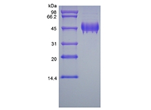
| Molecular Weight |
Approximately 44.4 KDa, a single non-glycosylated polypeptide chain containing 399 amino acids. |
| Biological Activity |
Fully biologically active when compared to standard. The ED50 as determined by its ability to enhance the adhesion of human Saos2 cells to bovine Collagen I coated plate is less than 2 ng/ml, corresponding to a specific activity of > 5.0 × 10⁵ IU/mg. |
| Appearance |
Sterile filtered white lyophilized (freeze-dried) powder. |
| Formulation |
Lyophilized from a 0.2 um filtered concentrated solution in 20 mM PB, pH 7.4, 150 mM NaCl. |
| Endotoxin |
Less than 1 EU/ug of rHuPEDF as determined by LAL method. |
| Reconstitution |
We recommend that this vial be briefly centrifuged prior to opening to bring the contents to the bottom. Reconstitute in sterile distilled water or aqueous buffer containing 0.1 % BSA to a concentration of 0.1-1.0 mg/mL. Stock solutions should be apportioned into working aliquots and stored at ≤ -20 °C. Further dilutions should be made in appropriate buffered solutions. |
| Stability and Storage |
Use a manual defrost freezer and avoid repeated freeze-thaw cycles.- 12 months from date of receipt, -20 to -70 °C as supplied.- 1 month, 2 to 8 °C under sterile conditions after reconstitution.- 3 months, -20 to -70 °C under sterile conditions after reconstitution. |
| References |
|
| SDS-PAGE |
 |
| Safety Data Sheet (SDS) Download |
Click to download |
| Technical Data Sheet (TDS) Download |
Click to download |