Interleukin-7 (IL-7) is encoded by the IL7 gene and secreted by stromal cells in the red marrow and thymus. It binds to the IL-7 receptor, a heterodimer consisting of IL-7 receptor alpha and IL-2 receptor gamma chain. IL-7 stimulates the differentiation of hematopoietic stem cells into lymphoid progenitor cells and also stimulates proliferation of B cells, T cells and NK cells. Murine IL-7 has approximately 65 % amino acid sequence identity with human IL-7 and both proteins exhibit cross-speciesactivity. IL-7 as an immunotherapy agent has been examinedin many human clinical trials for various malignancies and during HIV infection.
Source
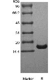
Escherichia coli.
Molecular Weight
Approximately 17.4 kDa, a single non-glycosylated polypeptide chain containing 152 amino acids.
Biological Activity
Assay #1:Fully biologically active when compared to standard. The ED50 as determined by a cell proliferation assay using murine 2E8 cells is less than 0.5 ng/ml, corresponding to a specific activity of > 2.0 × 10⁶ IU/mg.
Assay #2: Fully biologically active when compared to standard. The ED50 as determined by a cell proliferation assay using PHA-activated human peripheral blood lymphocytes (PBL) is 0.1-0.5 ng/mL. The specific activity of Recombinant Human IL-7 is approximately 2.0 × 10⁸ units/mg, which is calibrated against human IL-7 WHO Standard (NIBSC code: 90/530).
Appearance
Sterile filtered white lyophilized (freeze-dried) powder.
Formulation
Lyophilized from a 0.2 um filtered concentrated solution in PBS, pH 7.4.
Endotoxin
Less than 0.01 EU/ug of rHuIL-7 GMP as determined by LAL method.
Reconstitution
We recommend that this vial be briefly centrifuged prior to opening to bring the contents to the bottom. Reconstitute in sterile distilled water or aqueous buffer containing 0.1 % BSA to a concentration of 0.1-1.0 mg/mL. Stock solutions should be apportioned into working aliquots and stored at ≤ -20 °C. Further dilutions should be made in appropriate buffered solutions.
Stability and Storage
Use a manual defrost freezer and avoid repeated freeze-thaw cycles.A minimum of 12 months when stored at ≤ -20 °C as supplied. Refer to lot specific COA for the Use by Date.1 month, 2 to 8 °C under sterile conditions after reconstitution.3 months, -20 to -70 °C under sterile conditions after reconstitution.
References
1. Goodwin RG, Lupton S, Schmierer A, et al. 1989. Proc Natl Acad Sci U S A. 86:302-6.2. Sutherland GR, Baker E, Fernandez KE, et al. 1989. Hum Genet. 82:371-2.3. Lupton SD, Gimpel S, Jerzy R, et al. 1990. J Immunol. 144:3592-601.4. Noguchi M, Nakamura Y, Russell SM, et al. 1993. Science. 262:1877-80.5. Fry TJ, Mackall CL. 2002. Blood. 99:3892-904.6. Fry TJ, Mackall CL. 2002. J Hematother Stem Cell Res. 11:803-7.